2D-square ice crystals in a graphene sandwich
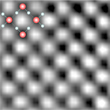
March 25, 2015 - Solid, liquid and gaseous, each child learns the phases of water in school. And also in science water is certainly one of the most studied substances. Nevertheless, researchers from the Universities of Ulm, Manchester and the University of Science and Technology of China were able to unravel a secret of the element: between two graphene layers they proved the existence of thin square ice crystals at room temperature (Fig. 1) [1].
Bulk water exists in many forms, including liquid, vapor and numerous crystalline and amorphous phases of ice, with hexagonal ice being responsible for the fascinating variety of snowflakes [2]. But water occurs at all interfaces, even in the driest desert, a microscopic layer covers all surfaces and can be detected in the smallest pores or is confined in cracks - for example in microscopic pores of rock. This very thin water is responsible for various phenomena in e.g. materials science, geology, biology, and nanotechnology [3-5]. Theory suggests many possible phases for adsorbed and confined water [6-11], but it has proved challenging to assess its crystal structure experimentally [12-15]. Furthermore, the theoretical results are sometimes very sensitive to modelled conditions and are appearing conflicting. For example, buckled monolayer ice [6] and flat hexagonal ice [9,11,15], respectively, were found inside hydrophilic and hydrophobic nanochannels below room temperature, whereas no in-plane order was observed for water inside simulated mica (hydrophilic) and graphite (hydrophobic) nanochannels at and above room temperature [8,10] A close analogue of planar square ice was seen in MD simulations of water inside carbon nanotubes [7,12], where water molecules form a monolayer that can be viewed as a sheet of square ice rolled up into a quasi-one dimensional cylinder. Neutron studies [12] revealed features consistent with the existence of such ‘ice nanotubes’, which melted above 50 K. Two dimensional (2D) ices have also been found experimentally on the surfaces of mica and graphite [13-15]. The studies using scanning probe microscopy [14] and electron crystallography [13,15] show that near-surface water can form correlated, solid-like layers. As for their intralayer structure, information is only available for 2D ices grown below 150 K, which were found to be hexagonal, with in-plane coordination similar to that of bulk ices [13,15].
For the TEM study of water between graphene, which was conducted in the frame of the SALVE project (here the low-voltage transmission electron microscopy for radiation-sensitive materials such as organic molecules will be developed further), the Ulm researchers Gerardo Algara-Siller and Ossi Lehtinen deposited a graphene monolayer on a standard transmission electron microscopy (TEM) grid, exposed it to a small amount of water and covered it with another graphene monolayer. Graphene is very well suited as substrate for molecules as its contribution to the image contrast can be eliminated by digitally filtering [16]. Furthermore, graphene’s mechanical strength, high thermal and high electrical conductivity and chemical stability protect to a certain degree radiation-sensitive materials against electron irradiation effects [16].
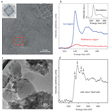
In the water/graphene sandwich experiments at Ulm University the majority of the samples remained intact during relatively long exposures to the electron beam (typically, 10–20 min); in a few cases only, they noticed etching of graphene [19]. Without graphene, frozen aqueous samples suffered from fast sublimation under the electron beam [17]. "In fact we first did not know what we were looking at when only observing the TEM images. Here the discussion with the Manchester group brought us to the right track, as they speculated about square ice already earlier. A detailed structural and elemental analysis then could remove any doubts: We saw square ice crystals," says Ute Kaiser, director of the group of EMMS at Ulm University (Fig. 2).
The transparent "graphene sandwich" used as a carrier here consists of two carbon monolayers, and the so-called van der Waals pressure between them caused the formation of this square ice structure at room temperature. Subsequently, the high resolution pictures were verified with image simulations (Fig. 3), which have beed performed by the University of Science and Technology of China. Two different models were used which yielded similar results. The pressure on the water molecules can be estimated on the basis of the adhesion energy for van der Waals materials [18]. Our collaborators in China found that the square crystals form if the pressure between the graphene layers is greater than 1 Gigapascal and the water film is thin enough - so they should occur in cracks in all materials at the nanoscale.
The structure of trapped water remained unknown up until now. "Microscopic cracks, pores or tiny channels are everywhere - and not only on our planet. For materials science, it is especially important to know that water behaves completely different on the nanoscale", says Irina Grigorieva, head of the research group in Manchester. This group also belongs to Sir Andre Geim, one of the scientists who received the Nobel Prize for their work on graphene in 2010. In view of the ultra fast water transport through graphene nano-channels, Geim had already speculated about square ice crystals - and the experiment has now confirmed him. There is still much to discover in the "nanoworld", he said. These results, recently published in the highly renowned scientific journal "Nature" [1], are important even beyond materials science and nanotechnology.
Algara-Siller, G., Lehtinen, O., Wang, F. C., Nair, R. R., Kaiser, U. A., Wu, H. A., Geim, A. K., & Grigorieva, I. V. (2014). Square ice in graphene nanocapillaries. Nature, 519: 443-445, doi: 10.1038/nature14295
Malenkov, G. (2009). Liquid water and ices: understanding the structure and physical properties. Journal of Physics: Condensed Matter, 21: 283101, doi: 10.1088/0953-8984/21/28/283101
Brown, G. E. (2001). How minerals react with water. Science, 294: 67-69, doi: 10.1126/science.1063544
Chandler, D. (2005). Interfaces and the driving force of hydrophobic assembly. Nature, 437: 640-647, doi: 10.1038/nature04162
Verdaguer, A., Sacha, G. M., Bluhm, H., & Salmeron, M. (2006). Molecular structure of water at interfaces: Wetting at the nanometer scale. Chemical reviews, 106: 1478-1510, doi: 10.1021/cr040376l
Zangi, R., & Mark, A. E. (2003). Monolayer ice. Physical review letters, 91: 025502, doi: 10.1103/PhysRevLett.91.025502
Takaiwa, D., Hatano, I., Koga, K., & Tanaka, H. (2008). Phase diagram of water in carbon nanotubes. Proceedings of the National Academy of Sciences, 105: 39-43, doi: 10.1073/pnas.0707917105
Giovambattista, N., Rossky, P. J., & Debenedetti, P. G. (2009). Phase transitions induced by nanoconfinement in liquid water. Physical review letters, 102: 050603, doi: 10.1103/PhysRevLett.102.050603
Han, S., Choi, M. Y., Kumar, P., & Stanley, H. E. (2010). Phase transitions in confined water nanofilms. Nature Physics, 6: 685-689, doi: 10.1038/nphys1708
Srivastava, R., Docherty, H., Singh, J. K., & Cummings, P. T. (2011). Phase transitions of water in graphite and mica pores. The Journal of Physical Chemistry C, 115: 12448-12457, doi: 10.1021/jp2003563
Bai, J., & Zeng, X. C. (2012). Polymorphism and polyamorphism in bilayer water confined to slit nanopore under high pressure. Proceedings of the National Academy of Sciences, 109: 21240-21245, doi: 10.1073/pnas.1213342110
Kolesnikov, A. I., Zanotti, J. M., Loong, C. K., Thiyagarajan, P., Moravsky, A. P., Loutfy, R. O., & Burnham, C. J. (2004). Anomalously soft dynamics of water in a nanotube: a revelation of nanoscale confinement. Physical review letters, 93: 035503, doi: 10.1103/PhysRevLett.93.035503
Yang, D. S., & Zewail, A. H. (2009). Ordered water structure at hydrophobic graphite interfaces observed by 4D, ultrafast electron crystallography. Proceedings of the National Academy of Sciences, 106: 4122-4126, doi: 10.1073/pnas.0812409106
Xu, K., Cao, P., & Heath, J. R. (2010). Graphene visualizes the first water adlayers on mica at ambient conditions. Science, 329: 1188-1191, doi: 10.1126/science.1192907
Kimmel, G. A., Matthiesen, J., Baer, M., Mundy, C. J., Petrik, N. G., Smith, R. S., Dohnálek, Z., & Kay, B. D. (2009). No confinement needed: Observation of a metastable hydrophobic wetting two-layer ice on graphene. Journal of the American Chemical Society, 131: 12838-12844, doi: 10.1021/ja904708f
Algara-Siller, G., Kurasch, S., Sedighi, M., Lehtinen, O., & Kaiser, U. (2013). The pristine atomic structure of MoS2 monolayer protected from electron radiation damage by graphene. Applied Physics Letters, 103: 203107, doi: 10.1063/1.4830036
Kobayashi, K., Koshino, M., & Suenaga, K. (2011). Atomically Resolved Images of I h Ice Single Crystals in the Solid Phase. Physical review letters, 106: 206101, doi: 10.1103/PhysRevLett.106.206101
Björkman, T., Gulans, A., Krasheninnikov, A. V., & Nieminen, R. M. (2012). van der Waals bonding in layered compounds from advanced density-functional first-principles calculations. Physical review letters, 108: 235502, doi: 10.1103/PhysRevLett.108.235502
Meyer, J. C., Eder, F., Kurasch, S., Skakalova, V., Kotakoski, J., Park, H. J., Roth, S., Chuvilin, A., Eyhusen, S., Benner, G., Krasheninnikov, A. V., & Kaiser, U. A. (2012). Accurate measurement of electron beam induced displacement cross sections for single-layer graphene. Physical review letters, 108: 196102, doi: 10.1103/PhysRevLett.108.196102
Näslund, L. Å., Edwards, D. C., Wernet, P., Bergmann, U., Ogasawara, H., Pettersson, L. G., Myneni, S., & Nilsson, A. (2005). X-ray absorption spectroscopy study of the hydrogen bond network in the bulk water of aqueous solutions. The Journal of Physical Chemistry A, 109: 5995-6002, doi: 10.1021/jp050413s


