SALVE I-II consultant Harald H. Rose at Ulm University
June 01, 2010
ULM—Harald H. Rose, the inventor of the practical realisable aberration correction and world leading expert in theoretical electron optics at Ulm University in the frame of the SALVE I-II project today. From February, 1st to May 31st, 2010 Ulm University ’s SFB 569 supported the SALVE I-II project with a guest scientist position for Harald Rose.
Rose's past, present and future: about his inventions in electron microscopy
Studies of Harald Rose hark back to a period of 30 years after the inventions of the electron microscope [1]. In 1959, he was a PhD student of Otto Scherzer who was a professor of Theoretical Physics at the University of Darmstadt, and performed his thesis investigating the imaging properties of arbitrary multipole systems. These studies were already aimed to find a feasible aberration corrector. In 1967 and 1971 [2, 3] Rose designed an improved spherical and chromatic aberration corrector that was basically technically functioning as could be demonstrated experimentally within the frame of the so-called Darmstadt Project in 1977 and 1982. Unfortunately, after the death of Otto Scherzer in 1982, the project was abandoned although it was successful as far as it went. In 1973, in collaboration with Plies, Rose designed an aberration corrector of complex systems with curved axes [4]. In 1982 he conducted a calculation of the magnetic dodecapol lenses [5].
In addition to advancing aberration correction, he made a number of other important contributions to electron microscopy. In 1978 he worked on the challenges associated with energy filters [6]. Decreasing the energy width of the electron beam reduces the contrast attenuation in the image. Rose dedicated himself not only to the development of electron optical components, he was inter alia engaged in the field of the theory of image formation by scattered electrons and particularly by inelastically scattered electrons in electron microscopy [7, 16]. This issue is of great importance in low voltage electron microscopy, since inelastic scattering increases at low voltages.
The spectral analysis opens the way to the investigation of chemical and electronic properties. Rose's approach to the analysis of the inelastically scattered electrons has been implemented practically in 1986. With his contribution, the design of a magnetic energy filter [8], the so-called omega-filter resulted for the 100 kV TEM at Fritz Haber Institute Berlin. Thereby an extremely sensitive alignment procedure enabled the successful operation of the Omega filter at high energy resolution (Figure 1).
The first aberration corrector, Zach and Rose designed for a scanning electron microscope in 1986 [9]. Surprisingly, even in 1990, electron microscopists in general were not convinced and declared the improvement in resolution by a hard ware corrector as "unthinkable" due to technical difficulties. In fact, the work of Rose in 1990 [10] enabled the breakthrough in aberration correction with his development of a technically feasible corrector. This aberration corrector was technically very easy through a combination of significantly simpler electron optical elements. It consisted only of one double-hexapole element with 4 additional, symmetrically placed lenses.
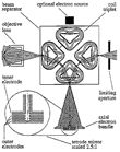
In 1992, together with Preikszas he developed an aberration corrector for a Low Energy Electron Microscope (LEEM) using a versatile beam splitter [11] that could subsequently be used in numerous devices (Fig. 2). Although the procedures for adjusting and controlling the alignment of spherically corrected electron microscopes required approximately 48 parameters to be set with an accuracy of 1:107 [12], they began to challenge the uncorrected instruments.
While the correction-development progresses, Rose was engaged in the development of a gauge invariance for the Eikonal method in the lens design in order to simplify the calculation of the required magnetic vector fields [13]. Plenty of challenges remained in the development of equipment to increase the energy resolution. 1994 Rose published an article, in which he described the Omega filter in detail from all theoretical and experimental aspects [14]. In 1995, together with Krahl he wrote also an article for Reimer's textbook "Energy filtered TEM" [15].
He also was active in the simulation of TEM images and contributed to a paper of C. Dinges that considered phonon and electronic excitations, that are an important part of the SALVE I-II project [16]. Rose’s inventions contributed to many different microscope techniques. In 1997, he developed an adaptation of his beam splitter in the SMART microscope [17].
Contrast delocalisation increases with spherical aberration. This causes imaging artefacts. Thust has shown in 1996 that these artefacts are a major impediment for the application of high-resolution instruments in atomically resolved defect and interfacial analysis [18]. In 1998, Rose, contributed to the work of M. Haider and K. W. Urban, that enabled the breakthrough for structure analysis; the application of hardware aberration correction in TEM [19, 20]. Contrast delocalization could, to a great extend, be avoided now and the interpretable resolution increased from 2.4 to 1.3 A in a 200 kV FEG-TEM.
Materials science on the atomic scale is absolutely dependent on the CS-correction and a first examination with a 2nd order-CS-corrected microscope could take place in 2002 [21]. Within the SATEM project, Benner et al have developed an aberration-corrected, monochromized electron microscope that contained the aberration corrector and the Omega filter proposed by Rose in 1986 in the projective system and combined sub-angstrom resolution with excellent options for analytical analysis [22].
The following publication by Rose in 2004 contained a description of this ultra-corrector that corrects all primary chromatic and geometrical aberrations [23]. One year later, Rose described the possibilities of aberrations-free electron microscopy as a unique tool for the investigation of materials at the atomic level [24]. Haider et. al. and the members of the company CEOS examined the practical realization of the combined CC / CS-corrector in medium voltage microscopes and reported their successful realization in 2008 [25]. Already in 2009, the corrector was used in the frame of the TEAM project.
In 2009 Rose described future trends in electron microscopy [27]. He included outlook and actual studies with phase plates, in which he had contributed, together with Barton and Benner, to Schroeder’s successful attempts to increase this way image contrast [26]. He discussed the specimen resolution as a function of instrumental and noise-related resolution. The latter is directly dependent on electron beam damage.
Worldwide Rose’s work is extremely highly evaluated, so for instance: Tang et. al. of Beijing National Laboratory for Condensed Matter, China say that [31] the aberration correction "achieved by Haider et al. following Rose’s idea [10], has attracted a great deal of attention among microscopists and materials scientists [19]". Taylor et. al. of Lawrence Berkeley National Laboratory, USA noticed that [32] "Ongoing efforts to correct spherical aberration of the objective lens [20] have been closely watched by the semiconductor industry with hopes that a new generation of CS-corrected microscopes might improve the current status of HR-TEM-based device characterization", Lichte et. al. from the field of electron holography, observed in 2007 [33] that "the CS-corrector [19] has resulted in enormous progress in all areas of TEM", and Malac, who like us, is also active in the field of imaging electron-beam sensitive materials, noted in 2008 [34] that "in the case of medium voltage microscopes, operating between about 50 and 400 kV, the 0.1 nm barrier was reached only recently thanks to the improved instrument stability and the development of spherical aberration correctors [19]".
Already in 2005, Ulm University has recognized the great potential of aberration correction and was able to order one of the first aberration corrected transmission electron microscope world-wide. The alignment was ordered not only at high voltages, where completely new insides into the nanoworld could be obtained, see e.g. [30]), but also, for the first time, at the lowest voltage of this microscope: 80kV, to obtain high resolution images of beam sensitive objects. (Interestingly, in 1970, Otto Scherzer had already pointed to the limitation of the actual resolution of an image by electron beam damage [28]). The image contrast especially at low voltages as well as the detailed functionality of the aberration corrector needed to be understood. Harald Rose was invited to a lecture series on his aberration correction for the research group of Electron Microscopy for Materials Science (EMMS) in 2006; this we can call also the very initial stage of the senior professorship of Harald Rose at Ulm University endowed by Carl Zeiss in the frame of the SALVE I-II project. "Through his whole theoretical and practical background and his outstanding numerous contributions to electron microscopy, Harald Rose matches exactly the challenging aims of the development of low voltage microscopy", Ute A. Kaiser, scientific leader of the SALVE I-II project, says. "We could not be more pleased. His new position combines the advantages of close and regular research contact between the SALVE I-II members at Ulm University with the exceptional possibilties for our students to gain inspiration, motivation and enthusiasm from a physicist, who is world leading expert in his field and has a lifelong teaching experience." Rose is enthusiastic about passing on his knowledge and experience to students at Ulm University and, moreover to the participants SALVE International School.
Selected publications (from 1932 to 2009)
- Knoll, M., and E. Ruska (1932): Das Elektronenmikroskop. Z. Phys., 78: 318-339
- ROSE, H. (1967), Über den sphärischen und den chromatischen Fehler unrunder Elektronenlinsen. Optik, 25: 587
- ROSE, H. (1971), Elektronenoptische Aplanate. Optik, 34: 285-311
- ROSE, H., and E. Plies (1973), Correction of aberrations in electron optical systems with curved axes. In: Image Processing and Computer-Aided Design in Electron Optic, (Academic Press, N. Y.): 344-369
- Haider, M., W. Bernhardt, and H. ROSE (1982), Design and test of an electronic and magnetic dodecapole lens. Optik, 63: 9-23
- ROSE, H. (1978), In: 9th International Congress on Electron Microscopy, 3: 230
- Kohl, H., H. ROSE (1985), Theory of image formation by inelastically scattered electrons in the electron microscopy. In: Adv. Electronics and Electron Physics, 65 (Academic Press, N. Y.): 173-227
- Lanio, and H. ROSE (1986): Test and improved design of a corrected imaging magnetic energy filter. Optik, 73: 56
- Zach J., and H. ROSE (1986), Efficient detection of secondary electrons in low voltage scanning electron microscopy. Scanning, 8: 285-293
- ROSE, H. (1990), Outline of a spherically corrected semi-aplanatic medium-voltage TEM. Optik, 85: 19-24
- ROSE, H., and D. Preikszas(1992), Outline of a versatile corrected LEEM. Optik, 92: 31-44
- Uhlemann S., M. Haider, and H. ROSE (1994), Procedures for adjusting and controlling the alignment of a spherically corrected electron microscope. ProcICEM-13 Paris, Les Editions de Physique, Paris I: 193-194
- Hoffstätter, G. H., and H. ROSE (1993), Gauge invariance in the eikonal method. In: Nuclear Instruments and Methods in Physics Research, A 328 (North Holland): 398-401
- ROSE, H. (1994), Correction of aberrations, a promising means for improving the spatial and energy resolution of energy-filtering electron microscopes. Ultramicroscopy, 56: 11-25
- ROSE, H., and D. Krahl (1995), In: L. Reimer (Ed.), Energy-filtering Transmission Electron Microscopy. (Springer Berlin): 43
- Dinges, C., A. Berger, and H. ROSE (1995), Simulation of TEM images considering phonon and electronic excitations.Ultramicroscopy., 60: 49-70
- Fink, R., M. R. Weiss, E. Umbach, D. Preikszas, H. ROSE, R. Spehr, P. Hartel, W. Engel, R. Degenhardt, R. Wichtendhal, H. Kuhlenbeck, W. Erlebach, K. Ihmann, R. Schlögl, H.-J. Freund, A. M. Bradshaw, G. Lilienkamp, T. Schmidt, E. Bauer, and G. Benner (1997), SMART: a planned ultrahigh-resolution spectromicroscope for Bessy II. J. Electron Spectrosc. Rel. Phenom., 84: 231-250
- Thust A., W. M. J. Coene, M. Op de Beeck, D. Van Dyck (1998), Focal-series reconstruction in HR-TEM: simulation studies on non-periodic objects. (1996) Ultramicroscopy, 64: 211-230
- Haider, M., S. Uhlemann, E. Schwan, H. ROSE, B. Kabius, and K. Urban (1998), Electron microscopy image enhanced. Nature, 392: 768-769
- Urban K., B. Kabius, M. Haider, and H. ROSE (1999), A way to higher resolution: spherical-aberration correction in a 200 kV transmission electron microscope. J. Electron. Microsc., 48: 821-826
- Kabius, B., M. Haider, S. Uhlemann, E. Schwan, K. Urban, and H. ROSE (2002), First application of a spherical-aberration corrected transmission electron microscopy in materials science. J. Electron. Microsc., 51: 51-58
- Weißbäcker, C., and H. ROSE (2002), Electrostatic correction of the chromatic and of the spherical aberration of charged-particle lenses II. J. Electron. Microsc., 51: 45-51
- ROSE, H. (2004), Outline of ultra-corrector compensating for all primary chromatic and geometrical aberrations of charged-particle lenses. In: Nuclear Instruments and Methods in Physics Research, A519: 12-27
- ROSE, H. (2005), Prospects for aberration-free electron microscopy. Ultramicroscopy, 103: 1-6
- Haider, M., H. Müller, S. Uhlemann, J. Zach, U. Loebau, and R. Hoeschen (2008), Prerequisites for a CC/CS-corrected ultrahigh-resolution TEM. Ultramicroscopy, 108: 1678-178
- Schroeder, R., B. Barton, H. ROSE, and G. Benner (2007), Contrast enhancement by anamorphotic phase plates in an aberration-corrected TEM. Microsc. Microanal., 13: 8-9
- ROSE, H. (2009), Future trends in electron microscopy. Phil. Trans. Roy. Soc. A – Math. Phys. Eng. Sci., 367: 3809-3823
- Scherzer, O. (1970), Die Strahlenschädigung der Objekte als Grenze der hochauflösenden Elektronenmikroskopie. Contributions of the Bunsen Foundation Phys. Chem., 74: 1154-1167
- Walther, T., E. Quandt, H. Stegmann, A. Thesen and G. Benner (2006), First experimental test of a new monochromated and aberration-corrected 200 kV field-emission scanning transmission electron microscope. Ultramicroscopy, 106: 963-969
- Zhang, Z. L., and U. A. Kaiser (2009), Structural imaging of b-Si3N4 by spherical aberration-corrected high-resolution transmission electron microscopy. Ultramicroscopy, 109: 9
- Tang, C. Y., J. H. Chen, H. W. Zandbergen, and F. H. Li (2006), Image deconvolution in spherical aberration-corrected high-resolution transmission electron microscopy. Ultramicroscopy, 106: 539-546
- Taylor, S. T., J. Mardinly, M. A. O'Keefe (2006), HR-TEM image simulations for the study of ultra-thin gate oxides. In: Scholarly publishing of the University of California: Lawrence Berkeley National Laboratory, 07-17-2001: 1-29
- Lichte, H., P. Formanek, A. Lenk, M. Linck, C. Matzeck, M. Lehmann, and P. Simon. (2007), Electron Holography: Applications to Materials Questions. Annual Review of Materials Research, 37: 539-588
- Malac, M., M. Beleggia, R. Egerton, and Y. Zhu (2008), Imaging of radiation-sensitive samples in transmission electron microscopes equipped with Zernike phase plates. Ultramicroscopy, 108: 2
Rose’s awards, honors and patents
Awards:
-
Honorary Professor of the Jiaotong University , Xian, China (since 1987)
-
Distinguished Scientist Award 2003 of the Microscopy Society of America
-
2005 Award of the 141 Committee of the Japanese Society for the Promotion of Sciences (JSPS)
-
Karl Heinz Beckurts Award 2006
-
Honda-Prize 2008 for Eco-technology
-
Richard Pohl Price 2009 of the German Foundation of Physics
-
Wolf Prize in Phyics 2011
-
BBVA Foundation Frontiers of Knowledge Award in Basic Sciences 2013
Honorary member of the
-
Microscopy Society of America
-
German Society of Electron Microscopy
-
Japanese Society for the Promotion of Sciences, and of the
-
Royal Microscopical Society
H. H. Rose is also the inventor of 105 patents on scientific instruments and electron optical components.


