SALVE project, start of 2nd phase
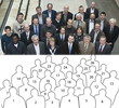
In February 2011, the SALVE Team celebrated the green light from the German Research Foundation (DFG) and the Baden Württemberg Ministry for Science, Research and Art (MWK) for the second period of the SALVE project (Fig. 1). To this event, the President of Ulm University, Professor Karl-Joachim Ebeling, welcomed the SALVE-Team and a top-class audience of future SALVE users at Ulm University. Professor Josef Zweck, Deputy Chairman of the German Society for Electron Microscopy (DGE) views SALVE as a door opener for the application of high-resolution transmission electron microscopy in biology, a branch which was considered as already being lost. Rasmus Schröder from Heidelberg University expressed his strong interest in the SALVE microscope and committed his further support with regard to cryo-microscopy at voltages lower than 60kV.
Ute A. Kaiser, the scientific leader of the SALVE project outlined during the ceremony (Fig. 2) the goals of the project, which comprise the exploration and development of low-voltage transmission electron microscopy for those radiation-sensitive materials, which are destroyed at higher acceleration voltages due to displacement of atoms by the incident electrons. She highlightened that the reduction of the accelerating voltage down to 20kV means to explore a largely undiscovered territory regarding microscopy modes, sample preparation, imaging theory, and the instrument itself. She presented a few key results of the first phase obtained by low-voltage aberration corrected electron microscopy 1 down to 20kV, obtained with the prototype SALVE I instrument, based on the LIBRA 200 from ZEISS NTS:
- Fullerenes @ double-walled carbon nanotubes have shown that the non-destructive examination time can be extended by a factor of about 100 by reducing the voltage from 80kV to 20kV (see Fig. 2 and [1]). This increase in tolerable dose significantly improves the attainable specimen resolution. The resolution of several organic and inorganic materials including very light atoms will thus benefit from using SALVE.
- A nitrogen defect in graphene can be imaged by its re-distribution of charge (see here on the SALVE website) and, moreover functionalizes graphene [2].
- A Cc corrector is mandatory for high contrast imaging at 20kV [3].
- A new approach for sample preparation by the focused ion beam technique has been developed resulting in ultra-thin (4nm for Si(111)) and unbent specimens ([1, 4]).
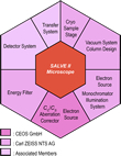
In the second phase of the cooperation project, the emphasis is on the development of a novel fully corrected electron microscope for low voltages (20-80kV). Kaiser further outlined the novel components of the low-voltage microscope (see Fig. 3) which have been evaluated during the feasibility phase. The heart of the new electron microscope is a Rose-Kuhn Cs-Cc-corrector [5, 6], modified and optimized for low voltages by CEOS. Criteria were less complexity with regard to construction, smallest length, and best optical quality. Further components of the SALVE microscope include a high-precision piezo-goniometer and a fast and efficient electron detector. Additional aspects of the work in SALVE II are the modification of existing imaging and spectroscopy methods and expanding the theory of contrast with slower electrons.
The lecture on aberration correction and its history was delivered by Maximilian Haider (CEOS GmbH, see Fig. 4).
Ute Kaiser further thanked her team member and consultant Carl-Zeiss Guest-Professor Harald H. Rose, for his enthusiasm and constant support (see Fig. 5). He is considered as the trendsetter in the field of aberration-correction and imaging theory. In 1990, Rose suggested a Cs-corrector type [7], that isn't limited by the famous Scherzer theorem proposed in 1936 [8], but based on a symmetric transfer doublet between two hexapoles, a very smart idea, which was indeed practical realizable as proven by Haider et al. in 1997 [9], opening a new area in physics: Imaging without aberrations in transmission electron microscopy. For their breakthrough, Harald H. Rose, Maximilian Haider together with Knut Urban were honored the Wolfprize for Physics in 2011.
Future SALVE users, the professors Tanja Weil (Institute for Organic Chemistry, Ulm University), Bernhard Keimer (Leibnitz-prize winner 2010, MPI-FKF Stuttgart) und Werner Tillmetz (Centre for Solar Energy and Hydrogene Research Baden Württemberg (ZSW) Ulm) expressed intentions to a future cooperation with the Group of Ute Kaiser at Ulm University in the fields of functionalized quantum dots, superconductors, and battery materials, respectively. Closing remarks delivered Klaus von Klitzing (Nobel prize winner 1986), who emphasized the cooperation in the field of graphene research.
First results of the project have been published in several journals (see [here] for a list of publications).
The SALVE project lasts five years. In the first phase the project financing was EUR 11.3 million and in the second phase further EUR 6.2 million. The SALVE microscope will be delivered to the University of Ulm in autumn 2013.
1: Obtained using the prototype SALVE I instrument, based on the LIBRA 200 from ZEISS NTS operated at 20 kV and the FEI Titan 80-300 instrument operated at 80 kV.
-
Kaiser, U. A. , J. Biskupek, J.C. Meyer, J. Leschner, L. Lechner, H. Rose, M. Stöger-Pollach, a N. Khlobystov, P. Hartel, H. Müller, M. Haider, S. Eyhusen, and G. Benner (2011), Transmission electron microscopy at 20kV for imaging and spectroscopy. Ultramicroscopy, 111: 1239-1246
Meyer, J. C., S. Kurasch, H. J. Park, V. Skakalova, D. Künzel, A. Groß, A. Chuvilin, G. Algara-Siller, S. Roth, T. Iwasaki, U. Starke, J. Smet, and U. Kaiser (2011), Experimental analysis of charge redistribution due to chemical bonding by High-resolution transmission electron microscopy. Nature Materials, 10: 209–215
Z. Lee, J. C. Meyer, H. Rose, U. A. Kaiser (2012), Optimum HRTEM image contrast at 20kV and 80kV – exemplified by graphene. Ultramicroscopy, 112: 39-46
L. Lechner, J. Biskupek, U. Kaiser (2011), Improved FIB target preparation of (S)TEM specimen - A method for obtaining ultra-thin lamellae. Microscopy and Microanalysis (publication and patent)
Rose, H. (1992), Proc. 10th Eur. Congr. El. Micr. (Granada, Spain), 47
Rose, H. (1990), Outline of a spherically corrected semi-aplanatic medium-voltage TEM. Optik, 85: 19-24
Scherzer, O (1936), Über einige Fehler von Elektronenlinsen. Z. Phys., 101: 593-603
M. Haider, S. Uhlemann, E. Schwan, H. H. Rose, B. Kabius, K. Urban, (1936), Electron microscopy image enhanced. Nature, 392: 768-769


