SHORT: New Insights into Damage Mechanisms in 2D c-MOFs at the Sub-Ångström Scale
March 01, 2024 - Knowledge of the atomic structure of layer-stacked two-dimensional conjugated metal–organic frameworks (2D c-MOFs) is an essential prerequisite for establishing their structure–property correlation. Scientists from Ulm and Nottingham University in Germany and UK have provided further detailed insights into the factors that enhance the stability of 2D c-MOFs in TEM, enabling the resolution of all structural features.
Recent years have witnessed a rise in the applications of two-dimensional layer-stacked conjugated metal−organic frameworks (2D c-MOFs), making effective use of their intrinsic conductivity, anisotropic charge transport, and (opto-)electronic properties [1]. Tremendous efforts have been devoted to band structure engineering of these emerging materials, which exhibit strong in-plane π-d conjugation, using the controlled assembly of transition metal nodes and aromatic ligands. This represents a challenging endeavor on both synthetic and characterization frontiers, with interfacial synthesis emerging as a new paradigm for the production of highly crystalline 2D c-MOFs [1]-[3].
Understanding the rational correlation between the electronic structure of 2D c-MOFs and the underlying atomic structure remains a formidable task that can be achieved through a combination of computational chemistry and imaging of these layer-stacked 2D structures with atomic resolution.
However, in aberration-corrected high-resolution transmission electron microscopy (AC-HRTEM), electron radiation damage, i.e., atomic displacement, bond scission, and chemical etching, can lead to instantaneous amorphization of organic materials during imaging [4],[5], severely limiting the achievable resolution. Additionally, as the thickness of the sample decreases from bulk toward the monolayer limit, the mechanism of the damage process needs to be revisited. Operating the AC-HRTEM in low-dose mode can be crucial to extract high-resolution information with minimal illumination [6].
For inorganic 2D materials, AC-HRTEM has demonstrated remarkable success in acquiring sub-angstrom resolution images [7],[8], and many experiments and calculations have been devoted to the quantitative determination of the damage cross section [9],[10]. High-quality monolayers of inorganic 2D materials are readily available, and their stability is high enough to acquire images with atomic resolution and to directly measure atomic positions with picometer precision, even in a defective crystal. Theoretical insights have been particularly helpful in further analysis of the damage process of inorganic 2D materials [11],[12]; however, this level of understanding of damage mechanisms in 2D c-MOFs is still lacking in the literature.
Understanding the Electron Stability of the c-MOFs.
To explain why the thin specimens of the studied structurally modified 2D c-MOFs show different stability under the e-beam, the authors suggest three main reasons: First, as expected, the replacement of the hydrogen−nitrogen fragments with oxygen increased the stability of the framework notably, by a factor of 2. This is due to the fact that hydrogen-containing bonds are weaker and get destroyed rapidly by the e-beam [13]. Also, once the hydrogen atoms are ejected from the structure, the associated nitrogen atoms become destabilized. Second, altering the density of copper centers in 2D c-MOF structures leads to a small difference in the critical dose. In the dense 2D c-MOF structures, the percentage of the damage with respect to the whole area remains the same as in the porous structure (two ejected atoms in the dense lattice result in the same percent damage as one atom in the porous lattice). The increase in atomic density may enhance the cage effect whereby knocked-out fragments are hindered and are unable to escape the sample, leading to recombination and self-healing of the lattice [14]. In the dense material, the fragments’ escape can be prevented. In the porous counterpart, however, channels are formed in the thicker crystal, and knocked-out fragments can escape through these channels, leading to a lower recombination rate and higher damage rate [15]. The biggest structure stabilization effect has been achieved by replacing the oxygen atoms with sulfur. Through this chemical substitutional change, the critical dose was increased nearly by a factor of 30, setting the stability of the Cu3(BHT) c-MOF far above the other 2D structures. This enormous change in the stability of Cu3(BHT) points to a fundamental difference in its physical properties.
Changing the structure of c-MOFs not only alters the chemical bonding but also can alter other properties that affect the stability of a c-MOF in the e-beam. For the Cu3(BHT) c-MOF, the electrical conductivity plays a major role. The here studied Cu3(HIB)2 has a conductivity of 13 S cm−1 [16], which is much higher compared to other structures like Cu3(HHB)2. Samples with higher conductivity have a greater potential to replace missing electrons or drain excess electron density, and the excited electrons have a faster relaxation rate; all of these processes significantly reduce radiolysis damage to the sample. However, due to the high hydrogen content, Cu3(HIB)2 is destroyed very quickly during imaging, and the conductivity of the material cannot compensate for the rapid destruction. The stability of Cu3(HIB)2 is comparable to the stability of the copper phthalocyanine (CuPc) self-assembly, which is a hydrogen-containing molecule, and the thin film of CuPc has the same order of conductivity as Cu3(HIB)2 [17]. As one of the most stable organic molecules, the dominating damage via displacement and radiolysis of bonds containing hydrogen leads to more than 1 order of magnitude lower stability than previous predictions [18].
A huge difference, however, was found for the Cu3(BHT) structure. Due to the strong π-d interactions and electron delocalization [19], Cu3(BHT) exhibits an electrical conductivity of 2500 S cm−1, which is by far the highest among 2D c-MOFs [20]. At the present stage, the conductivity of Cu3(BHT) is measured on a thin film, which might be affected by domain boundaries and defects [19]. In the TEM measurements of single crystalline domains, the electron conductivity could be even higher than the reported value. For typical c-MOFs, radiolysis is by far the largest contributor to e-beam damage, but not for the H-free Cu3(BHT). Here, the high conductivity suppresses radiolysis effects, and the remaining damage mechanism is knock-on damage.
Knock-on Damage of Cu3(BHT)
Due to the dominance of knock-on damage, the interaction between the e-beam and Cu3(BHT) is similar to that of inorganic 2D materials such as graphene and transition metal dichalcogenides (TMDs) [9],[10]. Computational analysis of the knock-on processes [11],[12] can be applied to describe the sample damage. In doing so, a vast array of intricate ejection processes has been detected in the Cu3(BHT) 2D c-MOF due to its structural complexity. To break down the damage cross section, the authors consider the computational cross section by primary knock-on atom (PKA) and by fragmentation energy; these ab initio molecular dynamics calculations have been done on a small Cu3(BHT) fragment as depicted in Figure 1B. Following a knock-on event, i.e., an electron hitting the PKA, different ejection processes have been identified, which depend on the energy transferred from an incident electron to the PKA. If the transferred energy is too small, no ejection occurs. As the transferred energy increases, the probability of ejecting a fragment goes up, and above a certain threshold, the primary ejection takes place in which the PKA is directly ejected on its own. Between these two limiting cases, the relaxation of the PKA during a knock-on event can cause sufficient structural disturbance to the sample for the process to be followed by the ejection of small fragments. The resulting intermediate fragmentation pathways consist of various small molecular particulates, which often include the PKA.
80 kV Cc/Cs Corrected Low-Dose HRTEM Imaging
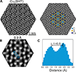
The outstanding stability of the Cu3(BHT) sample encouraged imaging at a lower electron accelerating voltage of 80 kV using the Sub-Angstrom Low-Voltage Electron Microscopy (SALVE) instrument (instrumental resolution is 0.78 Å) equipped with a chromatic (Cc) and spherical (Cs) aberration corrector. The Cc-correction converts the background noise induced by inelastic scattering into imaging signals, improving the S/N ratio and dose efficiency [21],[22]. However, sample stability is also a problem at these lower accelerating voltages. Since inelastic interaction is increased, radiolysis effects will increase at 80 kV compared to 300 kV. Figure 1A shows that at an accelerating voltage of 80 kV, knock-on damage can be largely avoided. Thus, high-resolution imaging of Cu3(BHT) at 80 kV is made possible by balancing both damaging processes. Due to the low scattering cross-section of carbon, unraveling the benzene rings in organic materials has been a long-standing challenge and is thus rarely reported [23]. The Cc/Cs correction combined with a reduced incident electron energy filter substantially increased the contrast in the HRTEM image, particularly at carbon sites (Figure 2A). Strikingly, the authors achieved an unprecedented resolution of 0.95 Å, enabling a clear distinction of neighboring carbon atoms (see Figure 2B and 2C).
Resource: Mücke, D., Cooley, I., Liang, B., Wang, Z., Park, S., Dong, R., Feng, X., Qi, H., Besley, E., & Kaiser, U. (2024). Understanding the electron beam resilience of two-dimensional conjugated metal−organic frameworks. Advanced Materials. DOI: 10.1002/adma.202406034
-
Wang, M., Dong, R., & Feng, X. (2021). Two-dimensional conjugated metal–organic frameworks (2D c-MOFs): chemistry and function for MOFtronics. Chemical Society Reviews, 50(4), 2764-2793. https://doi.org/10.1039/D0CS01160F
-
Liu, J., Chen, Y., Feng, X., & Dong, R. (2022). Conductive 2D conjugated metal–organic framework thin films: synthesis and functions for (opto‐)electronics. Small Structures, 3(5), 2100210. https://doi.org/10.1002/sstr.202100210
-
Yu, M., Dong, R., & Feng, X. (2020). Two-dimensional carbon-rich conjugated frameworks for electrochemical energy applications. Journal of the American Chemical Society, 142(30), 12903-12915. https://doi.org/10.1021/jacs.0c05130
-
Skowron, S. T., Chamberlain, T. W., Biskupek, J., Kaiser, U., Besley, E., & Khlobystov, A. N. (2017). Chemical reactions of molecules promoted and simultaneously imaged by the electron beam in transmission electron microscopy. Accounts of Chemical Research, 50(8), 1797-1807. https://doi.org/10.1021/acs.accounts.7b00203
-
Russo, C. J., & Egerton, R. F. (2019). Damage in electron cryomicroscopy: Lessons from biology for materials science. MRS Bulletin, 44(12), 935-941. https://doi.org/10.1557/mrs.2019.284
-
Qi, H., Sahabudeen, H., Liang, B., Položij, M., Addicoat, M. A., Gorelik, T. E., Hambsch, M., Mundszinger, M., Park, S., Lotsch, B. V., Mannsfeld, S. C., Zheng, Z., Dong, R., Heine, T., Feng, X., & Kaiser, U. (2020). Near–atomic-scale observation of grain boundaries in a layer-stacked two-dimensional polymer. Science Advances, 6(33), eabb5976. https://doi.org/10.1126/sciadv.abb5976
-
Linck, M., Hartel, P., Uhlemann, S., Kahl, F., Müller, H., Zach, J., Haider, M., Niestadt, M., Bischoff, M., Biskupek, J., Lee, Z., Lehnert, T., Börrnert, F., Rose, H., & Kaiser, U. (2016). Chromatic aberration correction for atomic resolution TEM imaging from 20 to 80 kV. Physical Review Letters, 117(7), 076101. https://doi.org/10.1103/PhysRevLett.117.076101
-
Haider, M., Uhlemann, S., Schwan, E., Rose, H., Kabius, B., & Urban, K. (1998). Electron microscopy image enhanced. Nature, 392(6678), 768-769. https://doi.org/10.1038/33823
-
Meyer, J. C., Eder, F., Kurasch, S., Skakalova, V., Kotakoski, J., Park, H. J., Roth, S., Chuvilin, A., Eyhusen, S., Benner, G., Krasheninnikov, A. V., & Kaiser, U. (2012). Accurate measurement of electron beam induced displacement cross sections for single-layer graphene. Physical Review Letters, 108(19), 196102. https://doi.org/10.1103/PhysRevLett.108.196102
-
Lehnert, T., Lehtinen, O., Algara–Siller, G., & Kaiser, U. (2017). Electron radiation damage mechanisms in 2D MoSe2. Applied Physics Letters, 110(3), 033106. https://doi.org/10.1063/1.4973809
-
Santana, A., Zobelli, A., Kotakoski, J., Chuvilin, A., & Bichoutskaia, E. (2013). Inclusion of radiation damage dynamics in high-resolution transmission electron microscopy image simulations: the example of graphene. Physical Review B, 87(9), 094110. https://doi.org/10.1103/PhysRevB.87.094110
-
Skowron, S. T., Lebedeva, I. V., Popov, A. M., & Bichoutskaia, E. (2013). Approaches to modelling irradiation-induced processes in transmission electron microscopy. Nanoscale, 5(15), 6677-6692. https://doi.org/10.1039/C3NR02130K
-
Chamberlain, T. W., Biskupek, J., Skowron, S. T., Bayliss, P. A., Bichoutskaia, E., Kaiser, U., & Khlobystov, A. N. (2015). Isotope substitution extends the lifetime of organic molecules in transmission electron microscopy. Small, 11(5), 622-629. https://doi.org/10.1002/smll.201402081
-
Egerton, R. F., Li, P., & Malac, M. (2004). Radiation damage in the TEM and SEM. Micron, 35(6), 399-409. https://doi.org/10.1016/j.micron.2004.02.003
-
Mücke, D., Linck, M., Guzzinati, G., Müller, H., Levin, B. D., Bammes, B. E., Brouwer, R. G., Jelezko, F., Qi, H., & Kaiser, U. (2023). Effect of self and extrinsic encapsulation on electron resilience of porous 2D polymer nanosheets. Micron, 174, 103525. https://doi.org/10.1016/j.micron.2023.103525
-
Dou, J. H., Sun, L., Ge, Y., Li, W., Hendon, C. H., Li, J., Gul, S., Yano, J., Stach, E. A., & Dincă, M. (2017). Signature of metallic behavior in the metal–organic frameworks M3(hexaiminobenzene)2 (M= Ni, Cu). Journal of the American Chemical Society, 139(39), 13608-13611. https://doi.org/10.1021/jacs.7b07234
-
Petersen, J. L., Schramm, C. S., Stojakovic, D. R., Hoffman, B. M., & Marks, T. J. (1977). A new class of highly conductive molecular solids: the partially oxidized phthalocyanines. Journal of the American Chemical Society, 99(1), 286-288. https://doi.org/10.1021/ja00443a070
-
Egerton, R. F. (2012). Mechanisms of radiation damage in beam‐sensitive specimens, for TEM accelerating voltages between 10 and 300 kV. Microscopy Research and Technique, 75(11), 1550-1556. https://doi.org/10.1002/jemt.22099
-
Huang, X., Sheng, P., Tu, Z., Zhang, F., Wang, J., Geng, H., Zou, Y., Di, C., Yi, Y., Sun, Y., Xu, W., & Zhu, D. (2015). A two-dimensional π–d conjugated coordination polymer with extremely high electrical conductivity and ambipolar transport behavior. Nature Communications, 6(1), 7408. https://doi.org/10.1038/ncomms8408
-
Huang, X., Zhang, S., Liu, L., Yu, L., Chen, G., Xu, W., & Zhu, D. (2018). Superconductivity in a copper(II)‐based coordination polymer with perfect Kagome structure. Angewandte Chemie, 130(1), 152-156. https://doi.org/10.1002/anie.201707568
-
Kabius, B., Hartel, P., Haider, M., Müller, H., Uhlemann, S., Loebau, U., Zach, J., & Rose, H. (2009). First application of Cc-corrected imaging for high-resolution and energy-filtered TEM. Journal of Electron Microscopy, 58(3), 147-155. https://doi.org/10.1093/jmicro/dfp021
-
Dunin-Borkowski, R. E., & Houben, L. (2016). Spherical and chromatic aberration correction for atomic-resolution liquid cell electron microscopy. In Liq. Cell Electron Microsc., 434-455. https://doi.org/10.1017/9781316337455.022
-
Zhang, D., Zhu, Y., Liu, L., Ying, X., Hsiung, C. E., Sougrat, R., Li, K., & Han, Y. (2018). Atomic-resolution transmission electron microscopy of electron beam–sensitive crystalline materials. Science, 359(6376), 675-679. https://doi.org/10.1126/science.aao0865


