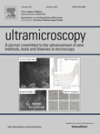
Ultramicroscopy highlights Low-Voltage Electron Microscopy by a special issue
October 12, 2014 - Ultramicroscopy, part of the international Elsevier group of science journals, edited by Angus I. Kirkland from Oxford Materials at the University of Oxford, is publishing a special issue of the journal with the topic Low-Voltage Electron Microscopy (LVEM) (Fig. 1). Here new results and future prospects of theory, instrumentation, applications, and general topics on LVEM are discussed. Ute A. Kaiser, director of the Sub Angstrom Low Voltage Electron microscopy (SALVE) project, and head of the Group of Electron Microscopy for Materials Science at Ulm University and Michael Stöger-Pollach from the Vienna University of Technology are guest editors of this issue of Ultramicroscopy.
Here in brief the content of this special issue: Three papers are dedicated to theory necessary to acquire and/or evaluate LVTEM data: from Zhongbo Lee et al. [1] (Ulm University), Jannik C. Meyer et al. [2] (University of Vienna) and Tatiana Latychevskaia et al. [3] (University of Zurich). New approaches for imaging electron-beam-sensitive materials by AC-TEM and other LVEM techniques are presented by Mitsuru Konno et al. [4] from Hitachi High-Technologies Corporation, Marian Mankos et al. [5] from Electron Optica Inc., and Takeo Sasaki et al. [6] of JEOL Ltd. Four other papers are dedicated predominantly to applications of low voltage techniques on different materials: David C. Bell et al. [7] (School of Engineering and Applied Sciences at Harvard University) report on practical materials science problems and show experimentally how different materials systems benefit from low voltage high resolution microscopy. Chris B. Boothroyd et al. [8] (Ernst Ruska-Centre Julich) report on imaging of barium atoms and functional groups on graphene oxide and Lawrence F. Drummy [9] (Air Force Research Laboratory, Wright Patterson Air Force Base) on organic–inorganic interfaces, and Jean-Nicolas Longchamp et al. [10] (University of Zurich) report on low-voltage electron holography. Finally fundamental questions on stability, resolving power, and the choice of accelerating voltage are addressed by Raymond F. Egerton [11] from Physics Department, University of Alberta, and Michael Stöger-Pollach [12, 13] from USTEM at Vienna University of Technology.
Analysis of our data base on aberration-corrected microscopy with respect to the potential of LV-AC-TEM
Today, LVEM is a topic of high relevance all over the world; productive research groups exist, and commercial companies are offering electron microscopes operating at accelerating voltages ≤ 80 kV (FEI Electron Optics, JEOL Ltd., Hitachi Ltd., & Nion Corp.). If we look back on just the last 8 years of global activities in low voltage AC-TEM, we see that this research field has constantly a very high percentage of publications in high impact factor journals. An evaluation of about 2000 publications showed that in low-voltage AC-TEM about 30% of the publications are published in journals with impact factor > 10 [‡], compared to only about 10% in medium voltage AC-TEM, illustrated in Fig. 2. This shows that the research of sensitive materials is of very high scientific interest. A more detailed statistical analysis of the contributions of LV-AC-TEM to science on the basis of the evaluation of publications in high-impact factor journals is shown in Fig. 3.
Despite the big global effort in imaging also biological objects with high resolution, only about 1.5% of the analyzed 2000 papers on aberration-corrected TEM are reporting on investigations of biological materials using AC-TEM [‡]. The statistic on these papers is presented in Fig. 4. Up until now, the absolute resolution limit is ~0.3 nm reported by Yu et al. 2011, they use a spherical aberration corrected FEI Titan Krios cryo electron microscope operated at 300 kV. Any clear improvement of this value would automatically increase studies of biological materials performed with aberration-corrected TEM and would imply a revolution in biology.
Richard P. Feynman has highlighted in his speech in 1959 the importance to image sensitive materials such as biological molecules and viruses in the TEM. Moreover Albert V. Crewe worked on imaging DNA back in 1971 and reported on problems concerning radiation damage. In his article "Radiation Damage: The Theoretical Background", Otto Scherzer stated: "Radiation damage puts a big question mark behind all efforts to observe the processes of life in an electron microscope and to image biomolecules at atomic resolution". Nowadays our community is still facing these challenges and strategies are developed to improve the situation.
In Germany a particular research project is even dedicated to unravel the atomic and electronic structure of radiation sensitive matter with 20 - 80 kV electrons; here academia and industry have come together and formed the SALVE project, funded by the German Research Foundation (DFG) and the Ministry of Science and Arts Baden Wurttemberg (MWK). The SALVE project began life in January 2009, with Ulm University as academic partner and FEI company and Corrected Electron Optical Systems GmbH (CEOS) as industrial partners. With Zeiss leaving the project in 2014 it will have a new industry partner, and the past activities are bearing fruit, not at least in a wealth of peer-reviewed scientific literature, about one third of it published in journals such as Science, the offsprings of Nature, or Nano Letters. Ute A. Kaiser says, "Over the past years, the crux of the matter have been identified, and we are quickly advancing towards our objectives. In five years, the SALVE project has produced more than 50 publications in major journals, one patent application and two prototype microscopes. We hope that the results are convincing to take hardware AC-LVEM from the instrument development state to the application in materials science and biology." In the SALVE project we are acutely aware that there is much work to do before AC-TEM becomes an established method for investigating beam-sensitive organic materials. In this special issue of Ultramicroscopy Harald H. Rose, academic supervisor of the publication by Lee et al. 2014 [1], has highlighted the importance of (1) the stability of the microscope and the microscope environment, (2) on the stability and suppression of noise in the chromatic and spherical aberration corrector. A news article about this publication on the SALVE website is available here. Furthermore new strategies for radiation damage reductions such as as enclosuring sensitive objects within carbon nanotubes or between graphene layers or isotope substitution need to be developed.
[‡] For evaluation we have used literature until the end of 2013, see the "Publication list for AC-TEM on the SALVE website". See also the pdf files of the "List of publications in journals with impact factor > 10 for AC-TEM." and "List of publications for AC-TEM of biological materials".
-
Lee, Z., Rose, H., Lehtinen, O., Biskupek, J., & Kaiser, U. (2014). Electron dose dependence of signal-to-noise ratio, atom contrast and resolution in transmission electron microscope images. Ultramicroscopy 145: 3-12, doi: 10.1016/j.ultramic.2014.01.010
-
Meyer, J. C., Kotakoski, J., & Mangler, C. (2014). Atomic structure from large-area, low-dose exposures of materials: A new route to circumvent radiation damage. Ultramicroscopy 145: 13-21, doi: 10.1016/j.ultramic.2013.11.010
-
Latychevskaia, T., Longchamp, J. N., Escher, C., & Fink, H. W. (2014). On artefact-free reconstruction of low-energy (30–250eV) electron holograms. Ultramicroscopy 145: 22-27, doi: 10.1016/j.ultramic.2013.11.012
-
Konno, M., Ogashiwa, T., Sunaoshi, T., Orai, Y., & Sato, M. (2014). Lattice imaging at an accelerating voltage of 30kV using an in-lens type cold field-emission scanning electron microscope. Ultramicroscopy 145: 28-35, doi: 10.1016/j.ultramic.2013.09.001
-
Mankos, M., Shadman, K., Persson, H. H. J., N’Diaye, A. T., Schmid, A. K., & Davis, R. W. (2014). A novel low energy electron microscope for DNA sequencing and surface analysis. Ultramicroscopy 145: 36-49, doi: 10.1016/j.ultramic.2014.01.007
-
Sasaki, T., Sawada, H., Hosokawa, F., Sato, Y., & Suenaga, K. (2014). Aberration-corrected STEM/TEM imaging at 15kV. Ultramicroscopy 145: 50-55, doi: 10.1016/j.ultramic.2014.04.006
-
Bell, D. C., Mankin, M., Day, R. W., & Erdman, N. (2014). Successful application of Low Voltage Electron Microscopy to practical materials problems. Ultramicroscopy 145: 56-65, doi: 10.1016/j.ultramic.2014.03.005
-
Boothroyd, C. B., Moreno, M. S., Duchamp, M., Kovács, A., Monge, N., Morales, G. M., Barbero, C. A., & Dunin-Borkowski, R. E. (2014). Atomic resolution imaging and spectroscopy of barium atoms and functional groups on graphene oxide. Ultramicroscopy 145: 66-73, doi: 10.1016/j.ultramic.2014.03.004
-
Drummy, L. F. (2014). Electron microscopy of organic–inorganic interfaces: Advantages of low voltage. Ultramicroscopy 145: 74-79, doi: 10.1016/j.ultramic.2014.05.001
-
Longchamp, J. N., Escher, C., Latychevskaia, T., & Fink, H. W. (2014). Low-energy electron holographic imaging of gold nanorods supported by ultraclean graphene. Ultramicroscopy 145: 80-84, doi: 10.1016/j.ultramic.2013.10.018
-
Egerton, R. F. (2014). Choice of operating voltage for a transmission electron microscope. Ultramicroscopy 145: 85-93, doi: 10.1016/j.ultramic.2013.10.019
-
Stöger-Pollach, M. (2014). A short note on how to convert a conventional analytical TEM into an analytical Low Voltage TEM. Ultramicroscopy 145: 94-97, doi: 10.1016/j.ultramic.2014.01.008
-
Stöger-Pollach, M. (2014). Low voltage EELS—How low? Ultramicroscopy 145: 98-104, doi: 10.1016/j.ultramic.2013.07.004


