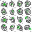
Endohedral metallo-fullerenes: AC-HRTEM allows accurate study of mechanism of structural transformations.
January 11, 2017 - The electron microscope is used not only to image chemical reactions with atomic resolution, but also to initiate these reactions. An international team of scientists has demonstrated the synthesis of new endohedral metallofullerenes (EMFs) within CNTs as well as imaging the dynamics of the structure development using the spherical aberration-corrected high-resolution electron microscope (HRTEM).
Endohedral metallofullerenes (EMFs) are fullerenes, which have an additional atom, ion or cluster in their interior. They have great potential for applications in biology and medicine and for molecular electronics. [1-3] The internal metal of an endohedral fullerene is effectively isolated from its surrounding environment, giving the endohedral fullerene a distinct advantage of high stability and low toxicity over the metal chelate complexes commonly used in radio-medicine and diagnostic radiology.
The traditional methods for the synthesis of EMFs via arc discharge reaction or laser evaporation are restricted to alkali earth elements, lanthanides and some transition metals with regard to the practically possible reaction products. The list of producible complexes can be expanded by a selected group of small molecules with modifications of the conventional methods of preparation [4, 5] such as the use of high pressure [6] or long sequences of organic reactions [7, 8]. However, numerous EMFs could not be manufactured.
The new method for the synthesis of EMFs is based on a procedure shown experimentally in the TEM by scientists from the Group of Electron Microscopy of Materials Science (EMMS) at the University of Ulm, Germany and their colleagues from the School of Chemistry at the University of Nottingham, UK. They demonstrated the conversion of graphene into fullerenes under electron irradiation [9]. Now they apply this method to encapsulate a cluster of other atoms. Such a method opens up possibilities to produce new types of EMFs by electron irradiation treatment of nanostructures starting from a metal cluster attached to sufficient carbon material, that is, about 100 carbon atoms.
“When the energy and dose rate of the e-beam are controlled, it is possible to trigger chemical reactions between metal clusters and carbon,” says Prof. Ute Kaiser, director of the SALVE project. “The formation of cage-like structures from amorphous carbon is driven by the energetic requirement of the carbon system to reduce the number of high energy, low coordinate atoms.”
The formation mechanism of the new nickel cluster EMFs achieved at room temperature is different from the previously proposed methods of metal−carbon nanostructure formation. The system is isolated, so that carbon supply from outside of the system is insignificant during the transformation. This suppresses carbon nanotube formation, thus enabling EMF formation. Moreover, the number of heavy metal atoms is conserved during the transformation. Thus, the EMFs produced with the new method give complete confidence of the composition of the final internal cluster.
Until now, EMFs could be synthesized at high temperature in the gas phase via the modified arc discharge method with a maximum of 4−7 atoms [7, 8]. By high-temperature treatment via laser irradiation multicage carbon nanoparticles with a metal core diameter of 5−10 nm could be formed. The new metallofullerene synthesis method fills the gap between these nano objects allowing cluster of up to 50−70 nickel atoms.
HRTEM imaging reveals important dynamics of metal−carbon bonding and a wide range of chemical processes catalyzed by metals under the influence of the kinetic energy of the e-beam have been observed with atomic resolution [10-14]. The AC-HRTEM used for this study is the image-side CS-corrected FEI Titan 80-300 transmission electron microscope of the Central Facility of Electron Microscopy at Ulm University. It was operated at 80 kV acceleration voltage with a modified filament extraction voltage for information limit enhancement and increased image contrast [15]. (see Fig. 2 and Video 1)
Video 1. Elemental carbon analyzed by AC-HRTEM imaging; carbon features extend from nickel clusters.
The time-dependent AC-HRTEM images clearly indicate that the nickel clusters act as a stable surface for the carbon cages to evolve, and provide a source of dissolved carbon atoms for the cage to grow (Fig. 2 a).
To extend the preparative reactions that lead to the new EMFs modeling and systematic comparison of processes under controlled e-beam irradiation and heating conditions is required. By molecular dynamics simulations [16], the separate role of electron irradiation and carbon nanotube templating within the formation of EMFs has been observed by comparing three specific cases of treatment of nickel clusters surrounded by amorphous carbon: electron irradiation inside a carbon nanotube, electron irradiation in vacuum, and heat treatment inside a carbon nanotube.
For electron beam irradiation in CNT (Fig. 3) the formation time was in the order of 1000 s and the accumulated dose of 4 × 10⁹ e−/nm2 correlates well with the characteristic time scales and doses observed in the experiments. Also the average inter-atomic distance between nickel atoms in the cluster of 2.4 Å is in good agreement with the experimental data.
The energy transmitted to the system from the electron beam is mostly transferred to the carbon atoms. This energy is almost exclusively spent on the transformation of the carbon nanostructure into the fullerene cage. In contrast, during heat treatment an important part of the energy is leading to cluster desorption and driving the metal through the forming fullerene cage. Thus, the electron irradiation is a required parameter to obtain high yield of EMFs. It is several times greater than under heat treatment.
The carbon nanotube acts as a very narrow container that prevents motion of the nickel cluster out of the fullerene cage and its subsequent desorption. But the yield of EMFs under electron irradiation inside carbon nanotubes is only slightly less than 50 % larger than that of the same numerical experiment in vacuum. Examples of structure evolution simulations that form the same nano-objects under electron irradiation in vacuum are shown in Fig. 4. The more perfect structure of EMFs formed in vacuum compared to the ones inside the nanotube is related to an increase in the accessible simulation time due to the decrease in size of the simulated system. The observations of carbon chains attached to the fullerene cage are also typical in simulations of fullerene formation. [17]
“This opens up possibilities for volume production of new types of EMFs and heterofullerenes in vacuum or buffer gas”, says Prof. Andrei Khlobystov from Nottingham University. “The process is analogous to the graphene flake to fullerene transformation under electron irradiation, which we have observed on graphite surface.”
The methods of synthesis of nanometer-sized graphene flakes are now actively elaborated [18]. Deposition of such flakes and metal clusters can be considered as one possible way to obtain EMFs and heterofullerenes under electron irradiation.
Challenges contain the prevention of merging of the forming EMFs or heterofullerenes. Because EMFs can easier diffuse on the surface after their formation in comparison with initial carbon−metal nanostructures, such diffusion can be used for their separation.
"With our comprehensive characterization and preparation opportunities we could again shed light on the mechanisms of the carbon shell formation around metal atoms and expand the range of synthesis in nano scale containers,” said Kaiser. “New electronically, optically and magnetically active inorganic nanostructures can possibly be produced in this manner in the future."
Resource: Sinitsa, A. S., Chamberlain, T. W., Zoberbier, T., Lebedeva, I. V., Popov, A. M., Knizhnik, A. A., McSweeney, R. L., Biskupek, J., Kaiser, U. A., & Khlobystov, A. N. (2017). Formation of nickel clusters wrapped in carbon cages: towards new endohedral metallofullerene synthesis. Nano Letters, 17: 1082–1089, doi: 10.1021/acs.nanolett.6b04607, [PDF], see also the supporting information, and Video 1, Video 2, Video 3.
Cong, H., Yu, B., Akasaka, T., & Lu, X. (2013). Endohedral metallofullerenes: An unconventional core–shell coordination union. Coordination Chemistry Reviews, 257: 2880-2898
Popov, A. A., Yang, S., & Dunsch, L. (2013). Endohedral fullerenes. Chemical Reviews, 113: 5989-6113.
Yang, S. (2014). Endohedral fullerenes: From fundamentals to applications. World Scientific, Singapore, 2014.
Stevenson, S., Rice, G., Glass, T., Harich, K., Cromer, F., Jordan, M. R., Craft, J., Hadju, E., Bible, R., Olmstead, M. M., Maitra, K., Fisher, A. J., Balch, A. L., & Dorn, H. C. (1999). Small-bandgap endohedral metallofullerenes in high yield and purity. Nature, 401: 55-57.
Wang, C. R., Kai, T., Tomiyama, T., Yoshida, T., Kobayashi, Y., Nishibori, E., Takata, M., Sakata, M., & Shinohara, H. (2001). A scandium carbide endohedral metallofullerene: (Sc2C2)@C84. Angewandte Chemie International Edition, 40: 397-399.
Saunders, M., Jiménez-Vázquez, H. A., Cross, R. J., & Poreda, R. J. (1993). Stable compounds of helium and neon: He C60 and Ne C60. Science, 259: 1428-1431.
Komatsu, K., Murata, M., & Murata, Y. (2005). Encapsulation of molecular hydrogen in fullerene C60 by organic synthesis. Science, 307: 238-240.
Kurotobi, K., & Murata, Y. (2011). A single molecule of water encapsulated in fullerene C60. Science, 333: 613-616.
Chuvilin, A., Kaiser, U., Bichoutskaia, E., Besley, N. A., & Khlobystov, A. N. (2010). Direct transformation of graphene to fullerene. Nature chemistry, 2: 450-453.
Helveg, S., López-Cartes, C., Sehested, J., Hansen, P. L., Clausen, B. S., Rostrup-Nielsen, J. R., Abild-Pedersen, F., & Nørskov, J. K. (2004). Atomic-scale imaging of carbon nanofibre growth. Nature, 427: 426-429.
Chamberlain, T. W., Meyer, J. C., Biskupek, J., Leschner, J., Santana, A., Besley, N. A., Bichoutskaia, E., Kaiser, U., & Khlobystov, A. N. (2011). Reactions of the inner surface of carbon nanotubes and nanoprotrusion processes imaged at the atomic scale. Nature chemistry, 3: 732-737.
Zoberbier, T., Chamberlain, T. W., Biskupek, J., Kuganathan, N., Eyhusen, S., Bichoutskaia, E., Kaiser, U., & Khlobystov, A. N. (2012). Interactions and reactions of transition metal clusters with the interior of single-walled carbon nanotubes imaged at the atomic scale. Journal of the American Chemical Society, 134: 3073-3079.
Koshino, M., Niimi, Y., Nakamura, E., Kataura, H., Okazaki, T., Suenaga, K., & Iijima, S. (2010). Analysis of the reactivity and selectivity of fullerene dimerization reactions at the atomic level. Nature chemistry, 2: 117-124.
Yue, Y., Yuchi, D., Guan, P., Xu, J., Guo, L., & Liu, J. (2016). Atomic scale observation of oxygen delivery during silver-oxygen nanoparticle catalysed oxidation of carbon nanotubes. Nature Communications, 7: 12251.
Biskupek, J., Hartel, P., Haider, M., & Kaiser, U. (2012). Effects of residual aberrations explored on single-walled carbon nanotubes. Ultramicroscopy, 116: 1-7.
Santana, A., Zobelli, A., Kotakoski, J., Chuvilin, A., Bichoutskaia, E. (2013). Inclusion of Radiation Damage Dynamics in High-Resolution Transmission Electron Microscopy Image Simulations: the Example of Graphene. Phys. Rev. B, 87: 094110.
Irle, S., Zheng, G., Wang, Z., & Morokuma, K. (2006). The C60 formation puzzle solved: QM/MD simulations reveal the shrinking hot giant road of the dynamic fullerene self-assembly mechanism. Journal of Physical Chemistry B, 110: 14531.
Bacon, M.; Bradley, S. J.; Nann, T. Graphene Quantum Dots. (2014) Part. Part. Syst. Charact., 31: 415−428.


